Dry (layered) blood cell analysis is a screening tool that involves pricking the tip of your finger and dabbing a single drop of blood onto a microscope slide 8 times. The blood is then left to dry in the open air for approximately 30 seconds and once dried it is placed under the microscope and magnified onto a high resolution TV monitor/screen. The approximate time to complete the dry (layered) blood cell analysis is 10-15 minutes and it is always done after the live blood cell analysis.
Live blood microscopy and dry blood analysis are two different diagnostic techniques used to assess a patient’s overall health status.
Live blood microscopy involves collecting a drop of blood from the patient’s fingertip, which is then examined under a microscope using darkfield, brightfield, and phase contrast microscopy. The blood cells are viewed in their natural, living state, providing real-time information about the condition and behavior of the cells. Live blood microscopy can reveal imbalances in the immune system, nutrient deficiencies, toxicity levels, and signs of oxidative stress.
On the other hand, dry blood analysis (also known as dry blood testing or layered blood analysis) involves collecting a drop of blood from the patient’s fingertip and allowing it to dry on a microscope slide. The dried blood is then examined under a microscope using darkfield, brightfield, and phase contrast microscopy. Dry blood analysis can reveal inflammation, degeneration, and toxicity of connective tissues and organs, as well as imbalances in the digestive and adrenal systems, magnesium deficiencies, heavy metal toxicity, and signs of oxidative stress.
In summary, live blood microscopy assesses the condition and behavior of blood cells in their natural state, while dry blood analysis evaluates the effects of blood cells on the body’s tissues and organs after they have dried.
During a live blood cell analysis session, you can expect the following:
- Consultation: The practitioner will first ask about your medical history and any current health concerns or symptoms. This will help them understand your health status and tailor the analysis to your specific needs.
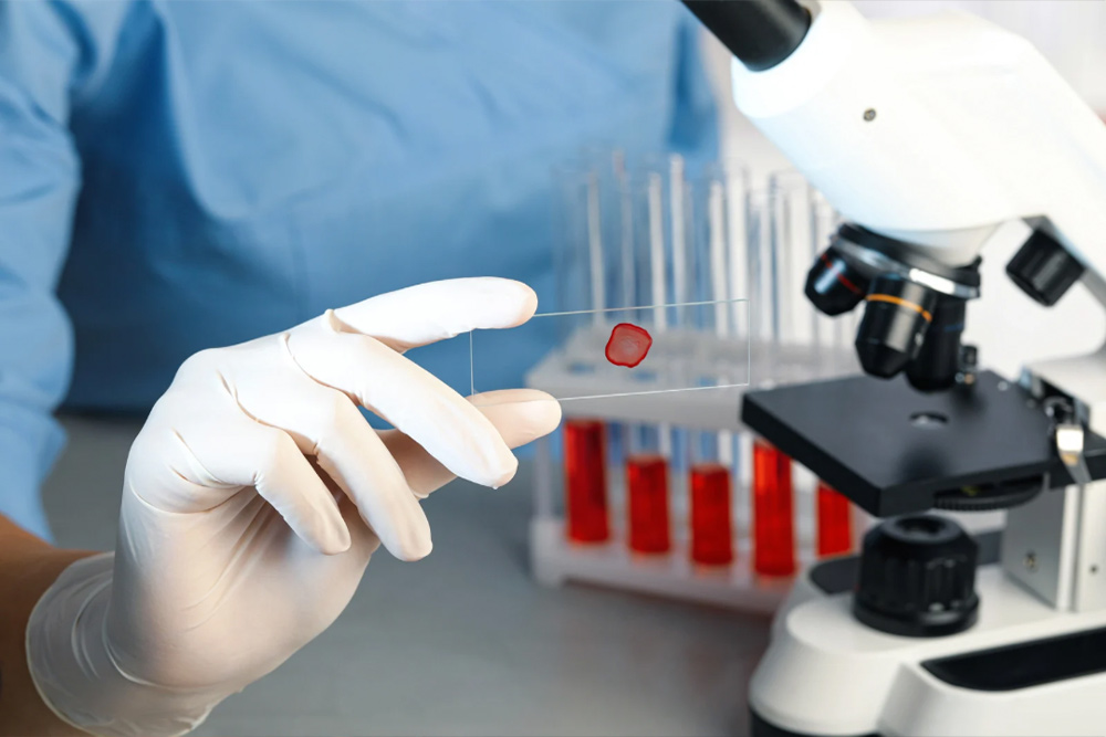
- Blood collection: The practitioner will then collect a small drop of blood from your fingertip using a lancet. The blood sample will be placed on a glass slide and immediately covered to prevent drying.
- Microscopy: The glass slide with your blood sample will be placed under a microscope, which may use darkfield, brightfield, or phase contrast microscopy. The practitioner will adjust the microscope settings to obtain a clear view of your blood cells.
- Analysis: As the practitioner views the live blood cells under the microscope, they will describe what they see and explain any abnormalities or imbalances. They may also take photos or videos of the blood cells for future reference.
- Recommendations: Based on the results of the live blood cell analysis, the practitioner may recommend lifestyle changes, dietary modifications, nutritional supplements, or other natural therapies to improve your health status. They may also refer you to a medical doctor for further evaluation or treatment if necessary.
Overall, a live blood cell analysis session usually takes around 30-60 minutes, depending on the practitioner’s approach and the complexity of your health status. It is generally painless and non-invasive, but you may experience a slight prick or discomfort when the blood sample is taken from your fingertip.